This post touches on the types of fungi that don't have the cap-and-stalk bodies that we associate with mushrooms. It's a category that includes, among others, brackets, sacs, cups, corals, clubs, and jellies. Remember, you can always click on a photo to view it in a larger format.
 |
| Jelly fungi. |
In my
previous post, I talked about "traditional" mushrooms, called boletes, that distribute their spores via pores on the underside of their caps. There is, in fact, another category of fungi with pores. They're called polypores. Most of them (the type discussed here) take shape as shelf-like protuberances from trees and are known as brackets. Unlike the quickly-decaying fruiting bodies of mushrooms, many brackets do not break down after spore distribution, instead building on layers year after year. While they may appear similar, not all polypores are closely related.
 |
| The brackets on this tree have been around long enough for moss to sprout on their tops. |
 |
| One of the most common kinds of polypores seen in my neck of the woods, growing on the end of a log. This particular specimen was about two inches across. |
 |
| You can see the white spores that have fallen from it on the moss below. |
 |
| You can just barely see the tiny pores here... |
 |
| …and clearly in this closeup imagine. |
 |
| This type of bracket closely mimicked the decaying bark on the surrounding logs... |
 |
| ….though bark doesn't usually grow on cut wood surfaces. |
 |
| Enlarged to show texture. |
 |
| My flash could just barely bring out the rough pores on the dark underside. |
 |
| These brackets, though fairly "woody" in texture, are slim and growing in a group... |
 |
| …unlike this solitary, bulky one. |
 |
| This chunky bracket is stone-gray in color... |
 |
| …while this leaner one bears stripes of autumnal orange. |
 |
| This big bracket (perhaps six inches in height) has been around a long time! Its lines and ridges are growth rings like those found on trees. |
 |
| Most of the "woody" brackets I saw have white pore surfaces, but this one is a sort of tan color... |
 |
| …and this pretty bracket... |
 |
| …has a delicately pink underside! |
 |
| You can see the texture created by the pores in this image. |
 |
| Other brackets are much more delicate-looking. |
 |
| They often had rippled, curving shapes. |
 |
| I noticed that the undersides of these brackets on the end of a log seemed very rough. |
 |
| The same was true of these ones I found on a log elevated enough that I could photograph the underside. |
 |
| I learned that jagged pore surfaces like these are considered "hydnoid," in that they resemble members of the hydnoid fungi group, otherwise known as tooth fungi. Some tooth fungi are, in fact, polypores, while some are not. |
Tooth fungi distribute their pores via hanging tooth- or spine-like points. Like many fungi categories, it was developed based on observation, creating a genus full of species that modern science has found to be only distantly related. However, since I am operating on a visual rather than a cellular level, the old definitions are sufficient for my purposes.
 |
| When I flipped over this stick to get a better look at the underside of this then new-to-me fungus, I found that it was quite rough in appearance. |
 |
| I've enhanced this photo to better show the tiny white "teeth" on this patch of fungi. |
 |
| I'm not sure where this lands on the hydnoid-polypore spectrum. |
 |
| Is it the same as the fungus on the branch? Is it Irpex lacteus, otherwise known as the milk-white toothed polypore? (It doesn't look exactly like any of the photos I found.) |
 |
| I DO know that it is white, has tubes (enlarged in the photo above), is rather ragged in appearance, and that I always found it on flat, vertical surfaces like the cut ends of logs. |
 |
| There is no doubt that THESE are in the tooth fungi family, however! The species is called Mucronella pulchra. |
 |
| I saw it in both white and yellow. Here some white specimens share a log with moss and mushrooms. Most of the formations I saw were very small, with the longest "teeth" not extending beyond a quarter of an inch, though the yellow ones above were a bit longer. |
Having talked about tooth fungi, which hang down, it's time to talk about clavarioid fungi, which point up, and particularly those simple, erect bodies referred to as club fungi. The same disclaimer--that these fungi are truly related only in appearance--that I've mentioned before applies to this group, too. They feed on decomposing wood and I've found quite a few of a particular member of the Xylaria genus both around my house and neighborhood and in the woods. These may very well be Xylaria hypoxylon, but lab tests would be needed to confirm it. The powdery white coating on the black "fingers" are asexual spores.
 |
| These "clubs" stand about two inches tall. |
 |
| This photograph and the one below are of the same grouping. |
 |
| As they matured, the clubs branched slightly. |
Speaking of branches… The branching members of the clavarioid group are known as corals because of their resemblance to aquatic coral. The kinds I found belong to the genus
Ramaria. They often grew in clumps, their delicate branches seldom more than two inches tall. I saw pink/peach, off-white, and purple/gray corals.
 |
| A beautiful peach coral fungus. |
 |
| A cluster of corals. |
 |
| The growth habit of this coral is quite convoluted! |
 |
| This off-white coral has elaborate branching. |
 |
| This smoky lilac coral is white at the tips. |
 |
| A coral sprouts from the same log as a group of gilled mushrooms. |
Next up: jelly fungi! This is what Wikipedia has to say about jelly fungi: "Jelly fungi are a paraphyletic group of several heterobasidiomycete fungal orders from different classes of the subphylum Agaricomycotina: Tremellales, Dacrymycetales, Auriculariales and Sebacinales. These fungi are so named because their foliose irregularly branched fruiting body is, or appears to be, the consistency of jelly." According to Canada's New Brunswick Museum, "The term 'jelly fungi' is an informal one applied to species of fungi having a gelatin-like consistency. The reason for this texture is that the structural hyphae of these fungi have walls that are not thin and rigid as they are in most fungi but instead are expanded out to a rather diffuse and indefinite extent...The peculiar texture of jelly fungi is not an absolute indicator of ancestral relationships." As you can see, especially in the first quote, if you really want to get into fungi identification, you also need to get very comfortable with a lot of clunky taxonomic vocabulary as well as some highly esoteric distinctions in mushroom morphology. From my strictly amateurish point of view, jelly fungi are cool because they are colorful and have bizarre forms. I found both witches butter (yellow) and purple jellydiscs in the woods.


One of the coolest things I found out in the woods were bird's nest fungi. I had not been aware such a thing even existed, so finding these tiny "nests" growing on fallen branches and other dead wood was very exciting. Bird's nest fungi consist of a fruiting body shaped very much like a nest that contains lenticular spore packages that look very much like eggs. Members of the Nidulariaceae family are "splash cups": that is, those little spore packages, called peridioles, are launched out of the "nest" when the "nest" sustains a direct hit by a raindrop. I found the whole thing quite magical--in a scientific way, of course!
 |
| A group of bird's nests on a fallen branch. They feed on decaying wood. |
 |
| Another group of bird's nest fungi. Membranes cover the openings of specimens that are not yet fully mature. |
 |
| The protective membrane of this bird's nest fungus has begun to split, revealing the "eggs" within. |
 |
| A group of mature bird's nests. |
 |
| A close-up look at the fringe-rimmed nests. Different types of bird's nest fungi have different colored peridioles. |
 |
| While the "splash cup" method of peridiole distribution can launch the little spore packets great distances, sometimes they only make it to the edge of the cup or just beyond... |
 |
| Here's a photo of my thumb next to a pair of bird's nest fungi to give you a proper sense of scale--they're about a quarter of an inch in diameter. |
Of all the strange fungi featured so far in this blogpost, puffballs (after brackets) are probably the most recognizable for the majority of people. Puffballs develop their spores within a closed cap with no gills or pores. When mature, the cap becomes brittle and eventually splits on its own or breaks under the pressure. (Raindrops can perform this function, as can curious small children.) The tiny spores are emitted in a smoke-like cloud, which gives puffballs their name.
 |
| A pair of maturing puffballs. |
 |
| A squirrel snacked on this very immature puffball. |
 |
| This mature puffball has broken open, revealing the body filled with spores. |
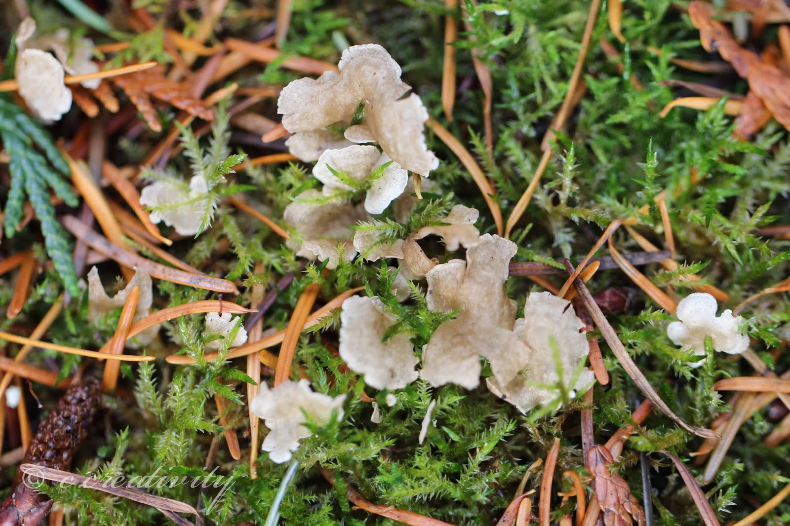
The fungus pictured below is known as
Arrhenia retiruga. I searched and searched and searched for an even a general ID for these tiny, pale, leafy forms that appeared below our birdbath back in October. For something so distinctive-looking, I could not, for the life of me, pull up anything that looked even close. The forms were somewhat reminiscent of leafy jelly fungi, but I knew that wasn't the right category: they weren't gelatinous enough and didn't grow in masses. I had actually given up on ID'ing them and considered my blogpost finished. I went outside to wipe my brain clean before a final proofreading and discovered that a new crop had sprouted around the birdbath--talk about fortuitous! I took more photos, dug up one of the specimens to see what it was growing on, and examined the uprooted specimen through my macro lens and a magnifying glass, trying to figure out where it fit in the fungi spectrum. It was the branching veins on the back that eventually led me to the identification; I happened to see a photo of a chanterelle with veining that was somewhat similar. That thought led me to the South Vancouver Island Mycological Society's "Key to Veined Fungi of the Pacific Northwest" and the key led me to
Arrhenia retiruga. I can't even begin to express the satisfaction of finally getting an ID after days and days of combing mushroom images! Like many other veined fungi, these are small members of a genera whose larger members have distinct gills, so while they are related to gilled mushrooms, they fit handily into this overview of fungi with neither caps nor stalks nor pores nor gills!
 |
| Convoluted, leafy, and scalloped in shape. |
 |
| The pale forms have subtle stripes or bands. |
 |
| They grew close together, but did not mass in clumps like leafy varieties of jelly fungi |
 |
| The first crop in October grew in a semicircular band, making me wonder if they were rooted on something underground. |
 |
| A new crop! |
 |
| The fungus was rooted in the moss by the finest filament. |
 |
| The underside of the fungus has a branching structure but no visible pores or gills. It was this branching that ultimately led me to make the identification. |
Every single mushroom pictured in this blog up this point--including my two previous posts on the subject--has been from the taxonomic division Basidomcycota. Despite the many forms they take, all members of the division share a type of spore-producing structure. The weird fungus below, however, comes from the division Ascomycota, called sac fungi because of the shape of their pore-producing structures. The one below is an elfin saddle fungus, Helvella vespertina, and is one of the coolest and weirdest fungi I've seen.
 |
| It stands about six inches tall. |
That sums up the third installment of the Fungi Files! There is more to come, including a second round of gilled mushroom photos and a look at lichens and molds, plus slime molds (which inhabit a category all their own), and photographs of bacteria colonies. If you missed the earlier post, you can view my gilled mushroom photos
here and my pore mushroom photos
here.


No comments:
Post a Comment